Is the Deadly Kissing Bug Disease the New HIV/AIDS?

photo credit: Wikipedia
Chagas, a disease caused by a parasite transmitted via the Triatoma bug (aka, the kissing bug), is claiming thousands of lives in Central and South America. Some experts are even calling it the “new HIV/AIDS of the Americas”. But is this comparison accurate, and how big a threat is the disease to the US?
Chagas is not a new disease. It’s named after Carlos Chagas (circa 1909), a Brazilian doctor who discovered that Triatoma carry a potentially lethal parasite called Trypanosoma cruzi. After several of his patients developed a strange infection that he couldn’t identify, Chagas set out to investigate how humans come in contact with the pathogen and its effects on the body of its host. He probably didn’t realize at the time that his work would prove to be groundbreaking in the history of epidemiology (Chagas later went on to identify the parasitic fungal genus Pneumocystis, another major discovery).
Kissing bugs, named because they bite the face and lips of humans (they’re also called assassin bugs), live in tropical climates near warm-blooded vertebrates to gain easy access to their blood. They stay hidden for much of the day, living in concealed places such as the cracks in a piece of wood and thatched roofs, and usually strike their victims as they sleep. The bugs defecate as they feed, allowing the parasite they carry to infect a new host.
According to the Centers for Disease Control and Prevention (CDC), the early stage of Chagas occurs immediately after infection and may have mild or no symptoms at all. Symptoms generally include fever, malaise, and a swelling of one eye. If left untreated, the infection can continue for years, often with no further symptoms, and over time it damages the heart, intestines and esophagus. There are two known treatments for the disease but they only work when administered in the early stages of infection. Once organ tissue has been damaged, it’s usually too late.
According to an editorial in the online journal PLoS, about 10 million people are currently living with Chagas, making it one of the most common neglected tropical diseases in Latin America and the Caribbean. It kills roughly 20,000 people worldwide every year. Lead author Dr. Peter Hotez, dean of the National School of Tropical Medicine at Baylor College of Medicine inHouston, warns that the disease is spreading globally and there may be between 300,000 and 1 million cases in the US.
While the infection is triggered by contact with a kissing bug, the disease appears to spread most rapidly via blood transfusions, organ transplants, and from mother to child during pregnancy — hence the comparison with HIV/AIDS.
There are, however, major differences between Chagas and HIV/AIDS, including that the parasite is not transmitted via sexual contact. And unlike HIV, which evaded detection in the blood supply for years, Chagas can be identified through blood screening and eliminated before it spreads. The problem is that screening requirements differ from country to country, and too often screening procedures are poorly administered or simply ignored.
Also unlike HIV/AIDS–at least in the early days of the disease–we know proven ways to prevent Chagas infection. Insect nets that prevent kissing bugs from biting humans are effective. Proper screening of the blood supply and reporting the disease are critical steps, as well as screening for the disease during pregnancy.
Is the disease a threat to the US? That depends on where you live. Texans, in particular, may have cause for concern because blood-donation screening is not mandatory in the state and physicians are not required to report the disease’s occurrence to health authorities, according to Wired.
What we do know is that Chagas is a major problem throughout Central and South America, and will continue to be until measures are taken to prevent its transmission. Unfortunately, those measures require adequate funding, and the countries most afflicted by the disease are also among the poorest. And since people can remain infected for up to 30 years before succumbing, we may have only started to see the true extent of the damage.
You can find me on Twitter @neuronarrative and at my website, The Daily Brain.
Chagas Disease: New AIDS Of The Americas Tropical Disease Brought By The Kissing Bug

image credit: trialx.com
Public Library of Science’s Neglected Tropical Diseases published an editorial in a medical journal
called the “The new AIDS of the Americas,” a tropical disease spread by insects called “chagas” similar to HIV/AIDS epidemic.
“There are a number of striking similarities between people living with Chagas disease and people living with HIV/AIDS,” the authors wrote, “particularly for those with HIV/AIDS who contracted the disease in the first two decades of the HIV/AIDS epidemic.”
Unlike HIV, Chagas is not a s-e-x-u-a-l-l-y transmitted disease: it’s “caused by parasites
transmitted to humans by blood-s-u-c-k-i-n-g insects.” It’s called the kissing bug. When it ingests your blood, it excretes the parasite at the same time. When you wake up and scratch the itch, the parasite moves into the wound and you’re infected.”
According to Dr. Peter Hotez, a researcher at Baylor College of Medicine and one of the
editorial’s authors, there are two problems:
First, once the heart symptoms start, which is the most dreaded complication—the Chagas
cardiomyopathy—the medicines no longer work very well and second the medicines are extremely toxic.
Apparently, more than 8 million people have been infected by Chagas, most of them in Latin and Central America. But more than 300,000 live in the United States.
Chagas, also known as American trypanosomiasis, k-i-l-l-s about 20,000 people per year, the journal said. Chagas disease is caused by blood s-u-c-k-i-n-g insects who transmit trypanosomes. watch the video.
The ‘new AIDS of the Americas’: Experts warn of deadly insect-borne disease that can cause victims’ hearts to explode
By Daily Mail Reporter
PUBLISHED: 20:13 GMT, 29 May 2012 | UPDATED: 08:49 GMT, 30 May 2012
A little-known life-threatening illness caused by blood sucking insects has been labelled the ‘new AIDS of the Americas’ by experts.
The parasitic illness called Chagas Disease has similarities to the early spread of HIV, according to a new study.
Similar to AIDS, Chagas is difficult to detect and it can take years for symptoms to emerge, according to experts writing in the journal PLOS Neglected Tropical Diseases.

The new AIDS? A parasitic illness called Chagas disease has been dubbed the ‘new AIDS of the Americas’ by experts. It is usually transmitted from the bite of blood-sucking insect species called Triatome bugs, which include ‘Kissing bugs’ (Triatomids)
An estimated 10 million people worldwide are infected with most sufferers in Bolivia, Mexico, Columbia and Central America, as well as approximately 30,000 people in the U.S., reported the New York Times.
The disease – once largely contained to Latin America – has spread into the U.S due to increases in travel and immigration.
THE DANGEROUS SPREAD OF CHAGAS DISEASE
Named after the Brazilian doctor who discovered it in 1909, Chagas disease is a potentially deadly illness spread by blood-sucking insects including Triatomids most commonly known as ‘kissing bugs.’
Like AIDS, the illness is difficult to detect and has a long remission period.
It spreads easily through blood transfusions and from mother to child.
Approximately a quarter of victims who contract the disease develop enlarged heart or intestines that can burst causing sudden death.
An estimated 10 million people worldwide are infected, including 30,000 people in the U.S.
Chagas is considered one of the Neglected Parasitic Infections, a group of five parasitic diseases that have been targeted by CDC for public health action.
It is estimated that in 2008 Chagas disease killed more than 10,000 people.
Due to the severity of the illness, the amount of people infected and the ability of prevention, Chagas is considered one of the Neglected Parasitic Infections, a group of five parasitic diseases that have been targeted by CDC for public health action.
Chagas commonly affects people in poverty-stricken areas and most U.S. cases are found in immigrants.
If caught early enough, the disease can be prevented with an intense 3-month drug treatment.
However, because of the lengthy incubation period and costly medication, Chagas is often left untreated.
Also known as the American trypanosomiasis, the disease spreads easily either through blood transfusions or, less commonly, from mother to child.
All blood banks in the U.S. and Latin America screen for traces of the disease.
Most blood banks in the U.S began screening for it in 2007.
Chagas is usually transmitted from the bite of blood-sucking insect species called Triatome bugs which release a parasite called Trypanosoma cruzi into the victim’s bloodstream.
The species includes Triatomids – black wingless beetles about 20mm in length commonly known as ‘kissing bugs’. Their closest relative is the Tsetse fly, found in Africa, which spread Sleeping Sickness (where the victim’s brain swells).
Chagas disease comes in two phases – acute and severe.
The acute phase may have no symptoms but can present a fever, general feeling of being unwell and swelling in one eye.

Victim: Charles Darwin may have contracted the disease as he travelled the globe on HMS Beagle in his 20s
After the acute phase the disease goes into remission and it can take years before symptoms, such as constipation, abdomen pain and digestive problems, emerge again in the severe stage.
The parasite can eventually make its way to the heart, where it can live and multiply.
About a quarter of the people who contract Chagas, develop enlarged heart or intestines that can burst causing sudden death.
Although the drugs available are not as expensive as those for AIDS, there are shortages of the medication in poorer countries and little money is being spent on discovering new treatments.
Chagas disease is named after Carlos Ribeiro Justiniano Chagas, a Brazilian doctor who first discovered the disease in 1909.
Researchers from the University of Maryland School of Medicine said last year that they believed Charles Darwin suffered from three different illnesses, including a Chagas infection.
The experts believe he contracted the disease during a five-year trip around the globe on the HMS Beagle in his 20s – and attributed it to his death of heart failure 47 years later.
The father of modern life scientists wrote in his journal that he had been bitten by a ‘wingless black bug’ during the expedition, where he visited South America.
_________________________________________
SOME OTHER BAD BUGS
Arthropod Vectors
Soft Ticks
Class: Arachnida
Subclass: Acari
Order: Ixodida
Family: Argasidae
Genus: Ornithodorous

Image 12-1. Ornithodoros – Soft Tick(SOURCE: CDC)

Image 12-2. Ornithodorus moubata, the most important soft tick disease vector found throughout the world (SOURCE: CDC)
General Characteristics
Soft Ticks are characterized by a tough leathery integument and a flattened oval shape when examined dorsally. The Argasidae lack the dorsal shield present in the Hard Ticks (Ixodidae), and need to be examined ventrally to observe their capitulum or mouthparts. Soft ticks generally have a world-wide distribution, with the most important disease vector Ornithodoros populatingEurope, Africa, Asia and the Americas.
Life Cycles

Illustration 12-1. Life Cycle of Soft Ticks. (SOURCE: Unknown)
Soft ticks have a hemimetabolous life cycle, with eggs hatching six legged larvae, which molt to eight legged nymphs. There are five to seven larval instars depending on the species, with each stage requiring a blood meal to proceed. Larvae of Ornithodoros moubata do not require a blood meal to molt to the nymph stage. Adult females lay small egg batches following each blood meal. The duration of the life cycle depends on the temperature, host availability, and inherently the particular species in question.
The adults are flat and oval in outline and have tough, leathery, wrinkled bodies. The mouthparts are situated underneath the body and are not visible from above. The eggs are laid in the places where the adults rest, such as cracks and crevices in the walls and floors of houses and in furniture. The larva, the five nymphal stages and the adults all actively search for hosts from which to take blood-meals. After feeding, which lasts about 30 minutes, they drop to the ground. Most species can survive for more than a year between blood-meals, and some for more than 10 years.
The soft ticks live apart from their hosts and are most common in the nests and resting places of the animals on which they feed. Some species, such as the chicken tick and the pigeon tick (Argasspecies) may feed on humans when the preferred hosts are not available.
Species that commonly feed on humans are found around villages and inside houses. Their habits are comparable to those of bedbugs: ticks often emerge from hiding places at night to suck the blood of humans and animals. Some species are common on travel routes, in rest houses and camping sites, and in caves and crevices.
Disease
Soft Ticks are vectors for serious disease including tick borne relapsing fever (Borrelia duttoni), rickettsial disease (Coxiella burneti), and some arboviruses. The most important disease spread by soft ticks is tick borne relapsing fever which occurs world-wide and is spread by spirochete infected Ornithodoros. Q-fever and arboviruses can be spreadfollowing a blood meal, but are both primarily introduced into the population by the Hard Ticks.
Tick-borne Relapsing Fever
This disease is caused by a microorganism of the genus Borrelia. It is transmitted by biting soft ticks of the genus Ornithodoros in many countries in the tropics and subtropics and also in Europe and North America. The ticks usually feed quickly at night in or near houses, and then leave the host.
The disease causes bouts of fever alternating with periods without fever. Death occurs in about 2-10% of persons who are untreated.
Treatment
Treatment is possible with tetracycline or its derivatives.
Prevention
Prevention requires measures to control soft ticks and to avoid their bites.
Hard Ticks
Class: Arachnida
Subclass: Acari
Order: Ixodida
Family: Ixodidae
Species:Ixodes, Amblyomma, Rhipicephalus, Dermacentor
General Characteristics
Hard ticks appear flattened when examined dorsally and can be primarily characterized by the presence of a dorsal plate or scutum, and a capitulum that projects beyond the body outline. The scutum regularly covers the entire dorsal area, although females may have a reduced plate present directly behind the capitulum. Ixodes spp. inhabit in Canada, Europe, Asiatic Russia, China, Japan, and Australia.


Image 12-3. Ixodes scapularis – male – showing the scutum covering the whole length of the body (left); Ixodes scapularis – female, showing the scutum covering half the length of the body (right)


Image 12-4. Amblyomma variegatum – male – showing the scutum covering the whole length of the body (left); Amblyomma variegatum – female – showing the scutum covering half the length of the body (right)

Image 12-5. Rhipicephalus sanguineus – female – showing the scutum covering half the length of the body (left); Rhipicephalus sanguineus – male – showing the scutum covering the whole length of the body (right).(AFPMB – used with permission)

Image 12-6. Dermacentor variabilis – male – showing the scutum covering the whole length of the body (left); Dermacentor variabilis – female – showing the scutum covering half the length of the body (right) (Image courtesy of: Iowa State University)
Life Cycle
Hard Ticks have a hemimetabolous life cycle similar to that of the Soft Ticks. Hard tick larvae search for suitable hosts, feed for several days, and drop off engorged to the ground below. Molting to the nymph stage follows several days later and the tick again searches for a suitable blood source. There is only one nymphal stage in Ixodes, and following several weeks of stasis the nymph will molt into an adult. Females lay one large mass of eggs, which forms a cellular mass on the scutum of the ovipositing female.
Disease
Hard ticks transmit a variety of diseases including Lyme Disease, Tick Paralysis, Rickettsia, arboviruses that are responsible for encephalitis and hemorrhagic fevers, tularemia and Babesia microtiinfection. Tick paralysis is caused by the toxins present in the saliva of the tick, and is introduced during feeding.
Lyme disease is a serious ailment caused by the spirochete Borrelia burgdorferi. Lyme disease results in acute erythema, systematic lesions and eventually chronic involvement of multiple organs.
Rickettsiae diseases spread by Ixodes include Q-fever (Coxiella burneti) and arboviruses that cause serious encephalitis.
Babesia microti infection primarily involves animals, with Man as an accidental host. The parasite is present in the tick salivary glands and is passed to Man via tick bite. Once in the bloodstream, the parasites enter the erythrocytes. Infection with B. microti ranges from asymptomatic to severe illness. Symptoms generally resemble those of malaria with fever, rigors, myalgia, and malaise. Occasionally there is mild or moderate hemolytic anemia and jaundice (renal failure). Parasitemia can be up to 25% in the immunocompetent individual, and up to 80% in patients who have been splenectomized.
Other Medically Important Hard Ticks
General Characteristics
Several other species of ticks are also responsible for the spread of disease throughout the world. Wood ticks (Dermacentor andersoni) are found in the mountainous west of North America; dog ticks (Rhipicephalus) are found in coastal areas. Lone Star ticks (Amblyomma americanum) are found in forests in SE USA where deer are found. The females of these species are known to cause a condition known as tick paralysis.

Image 12-7. Amblyomma, hard tick. The scutum is so-called ‘enameled’ colored areas, and is described as being an ornate species. (SOURCE: PHIL 5981 – CDC)

Image 12-8. Dermacentor variabilis, ticks of this species show very ornate scutums, covering the length of the body (left – male). When blood engorged it is harder to see the scutum (right – female). (SOURCE: PHIL 5977 – CDC)
Life Cycle

Illustration 12-2. One-host tick life cycle. (SOURCE: CDC)

Illustration 12-3. Two-host tick life cycle. (SOURCE: CDC)

Illustration 12-4. Three-host tick life cycle. (SOURCE: CDC)
Hard Ticks have a hemimetabolous life cycle similar to that of the soft ticks. Hard tick larvae search for suitable hosts feed for several days, and drop off engorged to the ground below. Molting to the nymph stage follows several days later and the tick again searches for a suitable blood source. There is only one nymphal stage in Ixodes, and following several weeks of stasis the nymph will molt into an adult. Females lay one large mass of eggs, which forms a cellular mass on the scutum of the ovipositing female.

Image 12-9. Female tick with egg mass. (SOURCE: PHIL 5968 – CDC)
Diseases
Both Dermacentor and Amblyomma transmit Rocky Mountain Spotted Fever (Rickettsia rickettsia). The different strains transmitted by these species vary in their virulence and subsequent manifestation of the disease. Dermacentor are known to transmit serious arboviruses responsible for encephalitis and hemorrhagic fevers. D. variabilis are also responsible for spreading tularemia (Francisella tularensis) and human granulocytic erlichiosis (Ehrlichia chaffeensis). Rhipicephalus sanguineus transmits Mediterranean Spotted Fever and African Tick Typhus.
Tick Paralysis
Hard ticks inject into the body with their saliva certain toxins that can cause a condition in people and animals called tick paralysis. It appears 5-7 days after a tick begins feeding, paralyzing the legs and affecting speaking ability, swallowing and breathing. It occurs worldwide and is most common and severe in children aged up to two years. Treatment involves removing the tick.
Tick-borne Rickettsial Fevers
This group of diseases is caused by closely related Rickettsia microorganisms transmitted by tick bites or contamination of the skin with crushed tissues or feces of the tick.
· Spotted fever due to Rickettsia rickettsii occurs in Brazil, Canada, Colombia, Mexico, Panama and the USA.
· Spotted fever due to R. sibirica occurs in Japan, the Russian Federation and the Pacific.
· Spotted fever due to R. conori is found in the Mediterranean region, Africa and southern Asia.
· Spotted fever due to R. australis occurs in Queensland, Australia.
· Q fever, caused by Coxiella burneti, has a worldwide distribution and is commonly present in abattoirs, meat-packing and meat-rendering plants, diagnostic laboratories, stockyards and poultry farms. It is transmitted to humans mainly by the consumption of milk and meat from contaminated cattle or the inhalation of dried infected tick feces by people working with cattle.
Symptoms in humans are sudden fever persisting for several weeks, malaise, muscle and joint pains, severe headache and chills. A rash sometimes spreads over the entire body. Death may result in about 15-20% of persons if the disease is misdiagnosed or left untreated.
Treatment
Antibiotics such as tetracycline or chloramphenicol can be used.
Prevention
Tick bites should be avoided and attached ticks should be removed rapidly and carefully. Several hours of attachment are needed before the Rickettsia organisms can infect humans.
Lyme Disease
Lyme disease (erythema chronicum migrans) is a severe and often debilitating condition caused by a spirochete, Borrelia burgdorferi. Acute Lyme disease is a flu-like illness, characterized by an expanding red rash in about 50% of patients, accompanied by fever, fatigue, and muscle and joint pain. Weeks or even months after the infecting tick bite, patients may experience swelling and pain in large joints (knee, elbow), encephalitis, facial palsy, ocular lesions and carditis, irrespective of whether a rash occurred in the acute phase. Later, perhaps years after the bite, there may be cartilage erosion (arthritis) and neuromuscular dysfunction (Fig. 4.29). Lyme disease occurs principally in northern temperate regions of the world, including China, Europe, the USA and the former USSR.

Illustration 12-5. A typical symptom of Lyme disease is swelling and pain in the large joints, such as the knees, and chronic arthritis. (SOURCE: Unknown)
Transmission
The disease is transmitted mostly by Ixodes ticks, commonly in the summer when the nymphs are abundant. Small rodents, especially mice, serve as reservoirs of infection while large mammals serve principally as hosts maintaining tick populations. The larvae acquire infection while feeding on mice, and nymphs or adults can transmit spirochetes during subsequent blood-meals. In the northern temperate zone, where it occurs most intensely, Lyme disease has become more common as deer populations have increased and as this critical host has adapted to living in closer proximity to people. In many areas, Lyme disease is acquired in the suburban residential environment.
Treatment
Further development of the disease in adults may be reduced or prevented by treatment with tetracycline or its derivatives for 2-4 weeks, and in children by treatment with penicillin.
Prevention
Prevention requires avoidance of tick habitats and bites, and vector control. Personal protection may be possible by the use of repellents on the skin and clothing in tick-infested areas. The removal of attached ticks within 24 hours may prevent spirochete transmission. Prophylactic antibiotic therapy may be desirable following the bite of an infected tick. New molecular assays are commercially available for detecting the spirochetes in tick samples.
Tularemia
Tularemia, also known as rabbit fever, deerfly fever and O’Hara disease, is caused by the infectious agent Francisella tularensis. The symptoms, which vary according to how the agent enters the body, include headache, chills, fever and the swelling of lymph nodes. The disease occurs in Europe, Japan, North America and the former USSR.
Transmission
Transmission takes place through the bites of ticks and deerflies (see Chapter 1) or as a result of handling infected animals such as rabbits and other game. Hunters and forest workers are at the highest risk of infection.
Treatment
Antibiotics such as streptomycin can be used to treat the disease.
Prevention
Tick bites and tick habitats should be avoided, impermeable gloves should be worn when skinning and dressing game animals, wild game meat should be thoroughly cooked, and untreated drinking-water should be avoided in areas where the disease occurs.
Tick-borne Viral Encephalitides
This is a group of viral diseases causing acute inflammation of the brain, spinal cord and meninges. The symptoms vary in severity with the type of disease. Many infections do not result in disease. Severe infections may cause violent headaches, high fever, nausea, coma and death.
· Far Eastern tick-borne encephalitis is found in the far east of the former USSR.
· Central European tick-borne encephalitis occurs in Europe from the Urals to France.
· Louping ill is a disease of sheep in the United Kingdom which sometimes affects people.
Transmission and Prevention
These diseases are transmitted by biting ticks and by the consumption of milk from infected animals. No specific treatment is available but vaccines have been developed against some of the diseases. Prevention requires avoidance or rapid removal of ticks.
Principal Hard Tick Vectors
Usually various tick species act as vectors for any one disease and their importance varies from region to region.
Disease
Vector
Lyme disease
Deer tick (Ixodes scapularis)
Spotted fever due to:
•
Rickettsia rickettsii
American dog tick (Dermacentor variabilis)
•
Rickettsia sibirica
Asiatic wood tick (Dermacentor silvarum)
•
Rickettsia conori
Brown dog tick (Rhipicephalus sanguineus)
•
Rickettsia australis
Wattle tick (Ixodes holocyclus)
Q fever
Lone star tick (Amblyomma americanum)
Tularemia
American rabbit tick (Haemaphysalis leporispalustris)
Far Eastern tick-borne encephalitis
Taiga tick (Ixodes persulcatus)
Central European tick-borne encephalitis
Castor bean ticks (Ixodes ricinus)
Kyasanur Forest disease
A tick of birds and monkeys (Haemaphysalis spinigera)
Colorado tick fever
American wood tick (Dermacentor andersoni)
Crimean-Congo hemorrhagic fever
A tick of birds and mammals (Hyalomma marginatum)
Table 12-1. Principle Hard Tick Vectors
Other Viral Diseases
Kyasanur Forest disease occurs in parts of India.
Omsk hemorrhagic fever is found in south-western Siberia; it causes severe disease and death in muskrat handlers; it is mainly waterborne, although it is found in hard ticks.
Colorado tick fever is a moderately severe disease that occurs in western North America.
Crimean-Congo hemorrhagic fever is an acute, often severe and fatal disease found in parts of Africa, Asia and Europe.
Mites
Class: Arachnida
Subclass: Acari
Order: Astigmata
Family: Pyroglyphidae
Genus: Dermatophagoides (Dust mites)
Family: Sarcoptidae
Genus:Sarcoptes (Scabies mites)
Order: Prostigmata
Family: Democidae
Genus:Demodex (Hair follicle mite)
Order: Mesostigmata
Genus: Leptotrombidium (Other Mites)
General Characteristics
Dermatophagoides, the common dust mite, commonly inhabit beds, mattresses, carpets and house dust. They are motile and feed on residual organic debris and are typically 0.3 µm in length. They characteristically have four long legs with suckers and pincer chelicerae.

Image 12-10. A Dust Mite (Dermatophagoides). They commonly inhabit mattresses, carpets and house dust.
Demodex folliculorum mites are 300 microns in length, live in the follicle for 5-6 days and migrate onto contiguous skin at night. Classically, they appear as 1 µm “sleeves” around the base of the eyelashes.

Image 12-11. Demodex folliculorum (SOURCE: CDC)
The mite, Sarcoptes scabiei, lives exclusively on human skin and causes scabies. It is small (the larger female is 0.3-0.4µm), colorless and oval with 8 short legs.

Image 12-12. Picture of the human parasite scabies (Sarcoptes scabiei). They live exclusively on human skin causing the condition known as scabies. (SOURCE: PHIL 3810 – CDC/Joe Miller/Reed and Crnrick pharmaceuticals.)
Leptotrombidium
adultsare normally reddish and covered with velvet like hair. It is the larvae that are parasitic and they too regularly appear reddish, but only have three pairs of legs. The palps are segmented, and the mouthparts are large and easily identifiable. Leptotrombidium larvae are distinguished by large eyes, two feathered sensillae and five feathered setae that are present on the scutum.

Image 12-13. Leptotrombidium (SOURCE: CDC of Taiwan)
Disease and Control
Dermatophagoides are antigenic, even when dead (fecal pellets are also allergenic), and are associated with complex allergies with symptoms such as asthma, perennial rhinitis, conjunctivitis and atopic dermatitis. A diagnosis can be made using reactive skin tests and treatment involves removal of accumulated antigens from mattresses and pillows.
Demodex have been implicatedin dermatitis, acne and should be considered in chronic, therapy-resistant cases of blepharitis. The role of this mite in ocular disease is uncertain. Washing with soap and water is the most effective method of prevention.
Symptoms of Sarcoptes infection arise after 4-6 weeks and comprise a papular pruritic eruption at a site often unrelated to the site of infestation; symptoms of secondary infestations arise almost immediately. Zoonotic scabies have shorter incubation periods but are also self-limiting. Persistent nodular scabies comprises itchy erythematous or scabbed nodules, often on the penis. When the immune system is impaired, Norwegian (crusted) scabies may develop: this presents with erythema and hyperkeratosis but little itching.
Diagnosis demands discovery of the mites, scybala or eggs by epidermal shave biopsy or superficial scraping. Burrows are best seen on wrists and interdigital spaces. They fluoresce under a Wood’s lamp after application of liquid tetracycline and, later, alcohol; alternatively ink may be used. Lindane (Quellada) lotion is the treatment of choice in the USA; permethrin or crotamiton are alternatives. Ivermectin has been recommended for Norwegian scabies. Malathion (Derbac-M) liquid or permethrin are recommended in the UK (benzyl benzoate is also active). Malathion should be avoided in infants and lindane should be avoided in pregnancy, breast-feeding and young children. Treatment is applied over the whole body except the head and neck and washed off after 24 hours. Normal laundering of bed linen and clothes is recommended. Household and sexual contacts should also be treated. Symptoms may continue after treatment because of persisting antigens, Calamine or Eurax lotion may be used; other problems include reinfestation and secondary bacterial infection.
Feeding by Leptotrobidium causes cellular damage and initially results in intense itching and irritation for the host. The agent of scrub typhus, Rickettsia tsutsugamuchi, is often transmitted by the larval trombiculid mite. Scrub typhus results in infection in those living or working near ‘mite islands’ which are usually found in transitional vegetation and provide a stable environment for mite proliferation. Prevention has generally been with chemical repellents or herbicides to remove the scrub habitat.
Mosquitoes
Class: Insecta
Order: Diptera
Family: Culicidae
Subfamily: Anophelinae
Genus: Anopheline
Subfamily: Culicinae
Genus: Culicine


Anopheles
(SOURCE: PHIL 7949 – CDC)
Aedes
(SOURCE: PHIL 9261 – CDC/ Prof. Frank Hadley Collins, Dir., Cntr. for Global Health and Infectious Diseases, Univ. of Notre Dame)
Culex
(SOURCE: PHIL 7957 – CDC)
Image 12-14. Anopheline and Culicine mosquitoes.
Introduction
Mosquitoes are small with a clearly demarcated body and very long slender legs. The head contains a large pair of kidney shaped compound eyes, a pair of antennae, and a single long proboscis for feeding. The thorax, abdomen and wings are often covered with scales. Differential coloration and pattern of these scales provides a means of visually distinguishing species. The large wings are folded over the segmented abdomen, which generally appears brown-black and slender but turns a bright red and swells following feeding.

Image 12-15.
Image of a typical Culicine mosquito. Adults are generally brown – black, at rest they place their abdomens parallel to the ground. (SOURCE: PHIL 4487 – CDC)
Mosquitoes may be classified as Anopheline (Anopheles) or Culicine (Aedes, Culex). The antennae of male mosquitoes are plumose (many feathery hairs); females are pilose (few spidery hairs). The male Anopheline palps are long and clubbed; those of the male Culicine are long but not clubbed. The female Anopheline palps are long; the female Culicine are short. If a mosquito is incorrectly sexed, a female Anopheline may be confused with a male Culicine.

Illustration 12-6. Image of typical male Anopheline mosquito showing the characteristic features of the mosquitoes head; the palps are as long as the proboscis with clubbed like ends and the antennae are plumose. The female Culicine mosquito shows the short palps and the pilose antennae. (SOURCE: CDC)
At rest, Anophelines usually position their abdomens at an angle to the surface; Culicines maintain their abdomen in a parallel position.

Illustration 12-7. Typical Anopheline and Culicine at rest, showing the abdomen at an angle to the surface. (SOURCE: CDC)
Anopheline (Anopheles) Mosquitoes
Class: Insecta
Order: Dipthera
Family: Culicidae
Subfamily: Anophelinae
Genus: Anopheline
Species: Anopheles

Image 12-16. Typical Anophelesmosquitoes (SOURCE: PHIL 7862 – CDC)
General Characteristics
Anopheles mosquitoes are characterized by dark and pale scale blocks arranged on their wings. They have palps that are of equal length to the proboscis, which appear terminally clubbed in males. Anopheles always rest at an angle when standing on surfaces and preferring to feed at twilight or night. Breeding sites are varied but Anopheles prefer unpolluted fresh or saltwater.
Life Cycle
Anopheles lay 50 to 200 dark color eggs in aquatic environments, and hatch in several days to several weeks depending on the external temperature. Anopheles larvae have a dark brown head and 6-7 anterior segments covered with dorsal palmate hairs. Accessory tegral plates are present on the dorsal side of segments 1-10 and two sets of anal papillae emerge from the last abdominal segment.
There are four larval instars that survive by filter feeding and breathing oxygen through their spiracles. Anopheles larvae occur throughout many different habitats including both permanent marshes and swamps, and temporary locations such as pots filled with water. In general Anopheles prefer to inhabit clean habitats. The larval period lasts about a week, but may be extended depending on the environmental conditions. The pupa is comma shaped with a set of trumpet shaped breathing tubes. The abdomen is covered with setae, and segments 2-7 have distinct spines. The pupal period may last a few days to weeks depending on the temperature.
Disease – Malaria
Anopheles are vectors of malaria, Bancroftian and Brugian filariasis and of multiple arboviruses (dengue fever; yellow fever; encephalitides and hemorrhagic fevers). Malaria is caused by Plasmodium falciparum, P. vivax, P. malariae and P. ovale. Transmission of the disease occurs in virtually all of tropical Africa, Central and South America, and the Middle and Far East. South East Asia is a particular problem due to multiple drug resistance. P. falciparum is found in Africa and other tropical countries as well as in subtropics. P. malariae has a low prevalence in both tropics and subtropics. P .vivax is the most widespread in temperate regions and subtropics but may also be found in the tropics. P. ovale has a low prevalence in West Africa. In Africa alone, 370 million people live in endemic areas.

Illustration 12-8. World map showing the distribution of malaria. (CDC)
P .vivax causes benign tertian malaria (43% of cases) and P. falciparum results in malignant tertian or sub-tertian malaria and pernicious malaria (50% of cases). P .ovale (mild tertian malaria, 1% of cases) and P. malariae (quartan malaria, 7% of cases) contribute a small percentage of malarial cases.
Clinical features including fever and chills are due to the host inflammatory response and are associated with rupture of erythrocytic schizonts. Fever presents in three stages – a) Cold: rigors and fever lasting 15 minutes to 1 hour; b) Hot: the patient is flushed with tachycardia and is pyrexial (40C) for 2-6 hours; c) Sweating: the temperature falls (over 2-4 hours). Each paroxysm lasts 8-12 hours in total. All erythrocytes containing a trophozoite will be destroyed within 48-72 hours. Periodic fever often takes more than seven days to develop, and anemia can be hemolytic or due to toxic marrow suppression. Splenomegaly occurs in all malaria: it may be acute or chronic (+/- hypersplenism). Jaundice may be hemolytic and/or hepatic (only P. falciparum). In addition, there may be headache, myalgia, arthralgia, diarrhea and vomiting.
Plasmodium falciparum is the most virulent form (invades mature and immature RBCs) and is often fatal if untreated. Blood schizogony takes place in deep capillaries and micro-circulatory failure can occur in individuals with little immunity to malaria. It does not relapse but recrudescence may occur. The time between paroxysms is 48 hours but fever may last for 24-36 hours. Very rapid progression and complications include diarrhea and vomiting; delirium; coma; convulsions; renal failure, including hemoglobinuria (blackwater fever); jaundice; pulmonary edema; hypoglycemia and abortion. Cerebral malaria often results in delirium, disorientation, stupor, coma, convulsions and death.
P. vivax / ovale exhibit 48 hours between paroxysms; relapses may occur up to 8 years after primary infection and only infects immature RBCs of those with Duffy blood group. Plasmodium malariaegenerally results in 72 hours between paroxysms, only infects older RBCs, and recrudescence may occur decades after primary infection.
The global malaria situation is serious and becoming worse: 300-500 million clinical cases occur annually. 1.5-2.7 million people die of malaria each year with approximately one million deaths among children under five years of age are attributed to malaria alone or in combination with other diseases. Countries in tropical Africa account for more than 90% of the total malaria incidence and the great majority of malaria deaths (WHO data). The death toll of African children with malaria is expected to double by 2010, conceivably reaching four million deaths per year. Many factors influence the epidemiology of this disease including: breeding habits of the various mosquito vectors; agricultural practices; economic conditions; industrialization and pesticide use. Increasing air-traffic from malaria endemic areas has led to the possibility of malaria developing in non-endemic areas where the mosquito vector has been imported onboard aircraft.
Treatments and Control – Malaria
If the infective species is not known, or the infection is known to be mixed, initial treatment should be with quinine, mefloquine or rarely halofantrine. Falciparum (malignant) malaria is often resistant to chloroquine and should be treated with quinine, mefloquine, halofantrine, quinidine or pyrimethamine-sulphadoxine. Benign malaria (P. vivax) should be treated with chloroquine although resistance has been reported from New Guinea. Malarial prophylaxis is relative and not absolute.
The UK Consensus Group on Malaria Prophylaxis (1997) recommend mefloquine for UK travelers to West, Central and East Africa for periods of greater than two weeks and for travelers to specific areas within south-east Asia: prophylaxis should be commenced two weeks before departure. Doxycycline can be used in older children and adults who cannot tolerate mefloquine.
Prevention is most dependent upon coverage of exposed skin and the use of insect repellent, mosquito nets impregnated with permethrin and correct prophylaxis. The vector may be controlled by water clearance programs, house spraying (DDT) and destruction of breeding areas. Drug resistance to DDT and ethical resistance to its use have limited its effectiveness. Natural immunity involves both antibody and cell-mediated systems and appears to require frequent boosting; antigens from different stages of the parasite’s life cycle will be important in vaccine development.
Disease – Filariasis
Anopheline mosquitoes also transmit the filarial worms Wuchereria bancrofti, Brugia malayi and Brugia timori. Wuchereria bancrofti is the main cause of “elephantiasis” (Bancroftian filariasis) and the most widely distributed filarial parasite of man. The adults live in the lymphatic system, and can survive for 30 years or more. Once they have mated they produce a pre-larval form, the microfilaria. Both the adults and the microfilaria may play a role in generating the symptoms and signs. Microfilaria measure 240-300mm in length by 7-10mm in width. They are sheathed (derived from ovum membrane) and nuclei terminate 15-20mm proximal to the pointed tail. There are fewer, more distinct nuclei than in other species and there are less body curves. Adult worms are slender and white (males 4cm; females stout and 10cm in length)
Initial infection with Wuchereria is usually asymptomatic. There may be recurrence of attacks of “cellulitis” affecting the limbs, breast, scrotum or elsewhere. Infection is associated with fever, lymphangitis, lymphadenopathy and occasionally abscess formation. These initially settle but later on the tissues eventually become edematous and hypertrophied. Further effects may include scrotal involvement and hydrocoele, which can lead to scrotal enlargement and lymph scrotum. This is “elephantiasis” and is associated with dermal hypertrophy, verrucous changes and the rupture of lymph varices into various sites.
Brugian (Malayan) filariasis is less widespread, less common and less serious than its Bancroftian counterpart. The life cycle is identical to that of Wuchereria bancrofti with Brugia malayi limited to Asia and B. timori restricted to Indonesia. Infection results in lymphadenopathy involving most frequently the inguinal area, lymphoedema normally below the knee, eosinophilia, and in rare cases chyluria.
Treatments and Control – Filariasis
Diethylcarbamazine (DEC) kills microfilaria. Ivermectin suppresses microfilaria production but its overall effectiveness remains untried and elephantiasis can be treated surgically. Control measures comprise draining of mosquito breeding sites and killing larvae. Many mosquitoes are resistant to insecticides but mosquito repellents and nets are effective. The infective pool may be reduced by periodic mass treatment with DEC. Brugia malayi is more susceptible to diethylcarbamazine (DEC) than is Wuchereria bancrofti. Anopheline larvae may be suffocated in their breeding sites but Culicine larvae (Mansonia sp.) derive oxygen from plants and are not vulnerable to such measures. Control depends upon the use of mosquito nets and periodic mass treatment.
Culicine(Aedes) Mosquitoes
Class: Insecta
Order: Dipthera
Family: Culicidae
Subfamily: Culicinae
Genus: Culicine
Species:Aedes
General Characteristics
Aedes can generally be distinguished by patterns of black and silvery scales present on the abdomen and thorax. The legs appear to have black and white rings along their length. The wings are generally covered with black scales. Aedes breed in marshes and other wetland areas and have a worldwide distribution.

Image 12-17. Typical Aedesmosquito. They are clearly distinguished from Anopheline mosquitoes due to the presence of black and slivery scales on the abdomen and thorax. (SOURCE: PHIL 9252 – CDC/ Prof. Frank Hadley Collins, Dir., Cntr. for Global Health and Infectious Diseases, Univ. of Notre Dame)
Life Cycle
Female Aedes lay eggs on damp areas such mud, detritus, clay and rock. The eggs are very robust and can survive desiccation and other environmental pressures. The eggs hatch in waves depending on the environmental cues. Aedes larvae have a stout barrel shaped siphon with one pair of subventral tufts. There are three pairs of setae on the ventral brush, and large setae are not present on the abdominal segments.
Disease
Aedes are vectors of Bancroftian filariasis and arboviruses such as yellow fever and dengue. Wuchereria bancrofti is the main cause of “elephantiasis” (Bancroftian filariasis) and the most widely distributed filarial parasite of Man. The adults live in the lymphatic system, and can survive for 30 years or more. They copulate and generate a pre-larval form, the microfilaria. Both the adults and the microfilaria may play a role in generating the symptoms and signs. Microfilaria measure 240-300mm in length by 7-10mm in width. They are sheathed (derived from ovum membrane) and nuclei terminate 15-20mm proximal to the pointed tail. There are fewer, more distinct nuclei than in other species and there are less body curves. Adult worms are slender and white (males 4cm; females, stout and 10cm in length).
Initial infection with Wuchereria is usually asymptomatic. There may be recurrence of attacks of “cellulitis” affecting the limbs, breast, scrotum or elsewhere. Infection is associated with fever, lymphangitis, lymphadenopathy and occasionally abscess formation. These initially settle but later on the tissues eventually become edematous and hypertrophied. Further effects may include scrotal involvement and hydrocoele, which can lead to scrotal enlargement and lymph scrotum. This is “elephantiasis” and is associated with dermal hypertrophy, verrucous changes and the rupture of lymph varices into various sites.
Yellow fever and dengue hemorrhagic fever are serious viral infections spread by the Aedes mosquito. Dengue is now the most important mosquito borne virus, with global infection increasing.
Control and Treatment
In general the most effective control for Culicine mosquitoes are repellents and fine screening or netting. Treatment with insecticides will also serve to reduce the vector population, but increased problems are encountered with Culicines because they also feed during the daytime. If filarial infection occurs, treatment with Diethylcarbamazine (DEC) will kill microfilaria. Ivermectin suppresses microfilaria production but its overall effectiveness remains untried and elephantiasis can be treated surgically.
Culicine (Culex) Mosquitoes
Class: Insecta
Order: Dipthera
Family: Culicidae
Subfamily: Culicinae
Genus: Culicine
Species: Culex
General Characteristics
Culex are distinguished by their lack of coloration and feature. The thorax, abdomen, legs and wings are often covered with brown-black scales giving a generally dark appearance. The abdomen may occasionally also have white scales arranged in segments. Culex breeds mainly in aquatic habitats, often in areas containing large quantities of organic waste.

Illustration 12-8. Typical illustration of a Culexmosquito, showing the characteristic brown-black scales on the thorax, abdomen, legs and wings giving it a general dark appearance. (Illustration: Trustees of the British Museum)

Image 12-18. ACulexmosquitotaking a blood meal from a human host. The abdomen becomes distended and blood red in color. (SOURCE: PHIL 4464 – CDC/Jim Gathany)
Life Cycle
Female Culex lay dark brown eggs in characteristic clumps of approximately 300 eggs. As mentioned these eggs are often found in organic waste deposits or polluted waters. Culex larvae have a long and narrow siphon with more than one pair of subventral tufts.
Disease
Culex mosquitoes are vectors of Bancroftian filariasis throughout Africa, but most importantly arboviruses such as Japanese encephalitis. Encephalitis occurs throughout the world, with Culex acting as an important vector for spread and infection. Culex mosquitoes are similar to Culicine mosquitoes, but prefer to bite at night and breed in organic refuse.
Control and Treatment
Culex mosquitoes are most easily controlled by improving sanitation and removing static water sources from the affected area. In general the most effective control for Culex mosquitoes are also repellents and fine screening or netting. Treatment with insecticides will also serve to reduce the vector population, but increased problems are encountered with Culicine mosquitoes because they also feed during the daytime. If filarial infection occurs treatment with Diethylcarbamazine (DEC) will kill microfilaria. Ivermectin suppresses microfilaria production but its overall effectiveness remains untried and elephantiasis can be treated surgically.
Tsetse Flies
Class: Insecta
Order: Diptera
Family: Glossinidae
Genus: Glossina
General Characteristics
Tsetse flies are large, yellow-brown or brown-black and measure 6-15µm in length. They are distinguished by a rigid projecting proboscis and a long pair of accompanying palps. There is a characteristic axe-shaped venation of the wings when viewed dorsally resembling an inverted hatchet in the central cell. The antennae appear short and feathery, and the abdomen is segmented and often striped or patched. Both males and females suck blood every 4-5 days, outside in open spaces.

Image 12-19. Typical illustration of a Tsetse Fly. They are yellow-brown in Colour and usually measure 6 – 15µm in length. Their characteristic feature is the hatchet shaped cell in the centre of the wing venation. (IMAGE SOURCE: Trustees of the British Museum)
Life Cycle
Female Tsetse flies are unique in the sense that they deposit larvae and do not lay eggs. The eggs mature within the female and are supplied with essential nourishment to complete larval development. This cycle requires a large number of blood meals to maintain and thus the female requires regular feeding. The larvae are normally deposited in shaded areas. There are three larval instars with the mature larva appearing white, visibly segmented with a pair of lobes at the posterior end. Pupation of the third instar results in a dark colored puparium with posterior lobes. The pupal period is extended (3-7 weeks) depending on the surrounding environmental conditions.
Disease
Tsetse flies are vectors for African sleeping sickness (Trypanosoma brucei sp. The two subspecies of T. brucei that infect Man are morphologically identical. T. b. gambiense causes Gambian sleeping sickness in Western Africa and T. b. rhodesiense causes Rhodesian sleeping sickness in East Africa. Another subspecies, T. b. brucei, causes nagana in cattle.

Illustration 12-9.
Distribution of African Trypanosomiasis throughout Africa (WHO, 1991)
Initially the patient has a headache, fever, chills and loss of appetite but specific clinical signs are absent. Parasitaemia comes in characteristic waves. Later, the spleen, liver and lymph nodes enlarge (Winterbottom’s sign). Finally, there may be CNS involvement leading to coma and death within several years. Trypanosoma brucei rhodesiense (rural East Africa): this is so acute that the patient invariably dies before classical symptoms develop. Without appropriate treatment, both forms are fatal.
Human African trypanosomiasis is rural and focal, with humans as the principal reservoir of infection of T. b. gambiense, and domestic cattle and wild animals as important reservoirs of T. b. rhodesiense. By the 1960s, it had been brought under control, but since 1970 the situation has deteriorated and the disease has reappeared, with major flare-ups in countries which have not maintained surveillance activities. It is estimated that 55-60 million people are exposed to the risk of becoming infected with trypanosomiasis, but only four million of them are under active surveillance or have access to health centers where reliable diagnosis is available: the estimated number of infected persons is over 300,000 (WHO data).
Treatment and Control
Anemia and other infections should be treated first. If given before the parasite has invaded the brain (hemolymphatic stage), suramin (Rhodesian disease) or pentamidine (Gambian disease) appear to be effective. For late disease (CNS involvement), drugs of choice are melarsoprol or eflornithine, with tryparsamide plus suramin as an alternative. Tsetse-infested areas should be avoided. Protective, light-colored clothing and repellents should be used. Tsetse flies are difficult to treat with insecticide (DDT has been tried) as their larvae live in burrows in the ground; protective vegetation must first be removed. Pentamidine prophylaxis is no longer advocated. Reservoir hosts should be identified and removed. It is possible to breed resistant or tolerant cattle (West Africa). Trypanosomes other thanT. brucei are also transmissible by Tabanid flies or by sexual contact.
Sand Flies
Class: Insecta
Order: Dipthera
Family: Psychodidae
Genera: Phlebotomus, Lutzomyia
Introduction
Sandflies are small (1.5-5µm) with a hairy head, thorax, antennae and wings. The antennae are long, may appear beaded, and protrude near a large set of black compound eyes. The wings are upwardly pointing at rest, and are a distinctive feature of Phlebotomine sandflies. Only the females are blood feeders, the males feed on plant nectar. Biting predominates nocturnally and they rest in moist and dark walls, cracks and tree trunks during the day.

Illustration 12-10. Typical illustration of a Sand Fly. The head, thorax, abdomen, legs and antennae are hairy, they are small usually measuring 1.5-5µm long. (Phlebotomus) (Illustration Source: Trustees of the British Museum)
Life Cycle
Female Phlebotomine sandflies deposit 30-70 minute eggs at each oviposition. The eggs are laid in dry areas, but require humidity to avoid desiccating. There are four instars and the mature larva is characterized by a distinct black head, 12 segments, thick bristles covering the body, and two pairs of caudal setae on their posterior end. As with other fly vectors the length of the larval stage depends on the ambient temperature, species in question, and food availability. The larval skin and caudal bristles remain attached at the posterior end during the pupal stage which lasts 5-10 days.
Disease
Sandflies are the only vectors for several species and subspecies of obligate intracellular protozoa responsible for leishmaniasis (Leishmania sp.) Cutaneous, mucocutaneous and visceral leishmaniasis are caused by different species of Leishmania contentiously linked to temperature preferences. Geographic location and host immune response also play a role in determining the form of disease.Leishmania tropica and L. major cause dermal cutaneous leishmaniasis; visceral leishmaniasis (kala-azar) is caused by L. donovani and mucocutaneous leishmaniasis is caused by L. braziliensis andL. mexicana. Visceral leishmaniasis in Europe is caused by L.infantum with dogs as the main reservoir. Leishmania tropica is found in the Middle East, North Africa, India and the Mediterranean. Leishmania donovani is found in the old and new worlds: South America, the Mediterranean, North and East Africa, India and China. Leishmania braziliensis is found in Central and South America. Leishmania mexicana is found in North and Central America, Texas and Mexico.
Dermal cutaneous leishmaniasis or Old World leishmaniasis is also known as “Tropical or Oriental Sore” or “Delhi Boil” and is generally localized to the skin surrounding the bite of the Phlebotomussandfly. The ulcers (volcano sign) are “draining” but produce “dry” ulcers that crust over. They usually resolve within one year but superinfection (yaws or myiasis) may occur and immunity develops. Cutaneous leishmaniasis affects over 300,000 people. The mucosal form affects the mucosa of the nose, pharynx, palate, larynx and upper lip causing ulcers that often become secondarily infected. Scarring can lead to death from pneumonia.
Visceral leishmaniasis, or “Kala-azar” is transmitted by Phlebotomus sandflies and is spread via the lymphatics from an often minor cutaneous lesion. They multiply in macrophages to form Leishman-Donovan bodies. Symptoms are usually chronic and comprise malaise, lymphadenopathy, cough, diarrhea, wasting and anemia, bleeding, and low-grade fever (3/day); liver and spleen enlarge and visibly distend the abdomen. Untreated, death ensues within three years, usually from secondary infection.
Leishmaniasis currently affects some 12 million people in 88 countries, all but 16 of which are in the developing world. It is estimated that 350 million people are exposed to the risk of infection by the different species of Leishmania parasite. The annual incidence of new cases is about two million (1.5 million of cutaneous leishmaniasis, and 0.5 million of visceral leishmaniasis). Recently, the WHO has reported an increase in overlapping of visceral leishmaniasis (VL) and HIV infection due to the spread of the AIDS pandemic. Leishmania / HIV co-infection is considered to be a real “emerging disease”, especially in southern Europe, where 25-70% of adult VL cases are related to HIV infection, and 1.5-9.5% of AIDS cases suffer from newly acquired or reactivated VL. Intravenous drug users have been identified as the main population at risk.
Treatment and Control
Dermal leishmaniasis usually resolves spontaneously. Visceral leishmaniasis is treated with extended courses of antimonial compounds (e.g. sodium stibogluconate or meglumine antimonate) and dietary supplementation (pentamidine isethionate has been used in antimony-resistant cases). WHO regimes are changing and pentamidine may be used. Mucocutaneous leishmaniasis is also treated with antimonial compounds. However, only around 50% of patients respond to antimonial compounds and relapses are seen. Pentamidine isethionate, paromomycin (aminosidine), allopurinol, ketoconazole, itraconazole, interferon gamma and liposomal amphotericin B have all been used; amphotericin B appears the most effective. Control of sandflies is difficult although buildings may be sprayed with insecticide. Sandflies cannot bite through clothing. Repellents are effective although mosquito nets are of limited value (sandflies are 3 µm). Sandflies are nocturnal and can be avoided. Rodent and dog control is a possibility. Vaccines (killed or live attenuated promastigotes) have been tried but effectiveness has not been assessed.
Black Flies
Class: Insecta
Order: Diptera
Family: Simuliidae
Genus: Simulium
Introduction
Black Flies are small (1.5-4µm in length) and normally black with short hairless legs and antennae. They have large compound eyes and a characteristically hairy humped thorax. Flies of the genusSimulium are generally found near free-flowing well-oxygenated water and bite during the day, tearing the skin to reach blood vessels.

Illustration 12-11. Typical illustration of a Black Fly(Simulium). They are usually small (1.4–4µm in length), black with short hairless legs and antennae.(SOURCE: PHIL 4639 – CDC)

Image 12-20. Image of a Black fly receiving a blood meal. (SOURCE: Unknown)
Life Cycle
Simuliidae eggs are laid in flowing or turbid waters in clusters of 200-800 eggs depending on the species. Hatching is mediated by the external environment, generally occurring several days following oviposition. There are 6-9 larval instars, with the larvae remaining essentially sedentary for the period and feeding through filtration. Movement is possible if necessary with some larval species acting as predators. The mature larvae can be recognized by a characteristic black “gill spot” on the thorax. The pupa is enclosed in a dark colored cocoon, identifiable by a series of filamentous respiratory gills protruding from the anterior end. In most cases the adult fly emerges from the pupal stage following one week of incubation.
Disease
Simulium are vectors for the filarial parasitic nematode responsible for Onchocerciasis (Onchocerca volvulus). It is distributed throughout Africa, Arabia, Central America, northern South America and Mexico with 30 million people infected in Africa alone; it is one of the most important causes of blindness in the world. Adult worms (up to 50cm) live in the subcutaneous tissue of man (the only known host) and release microfilariae into the skin. These are taken up by Simulium (S. damnosum and S. naevei in Africa; S. ochraceum and S. metallicum in Central America). Microfilariae penetrate the gut wall, migrate to the thoracic muscles, molt to L2 stages and molt again to filariform L3 stages which are passed to the next host during a blood meal. Larvae mature to adults in one year and may live for up to 20 years. Microfilariae can live for up to two years but are often trapped, in fibrous nodules, by the host’s cellular response and are responsible for the pathology. Microfilariae are highly motile, unsheathed and measure 300 by 7 microns; there are no nuclei in the end of the tail, which is long and pointed; the head is slightly enlarged. Microfilariae are not found in blood.
Skin problems include nodules (2cm to 5cm in diameter) containing adult worms. Onchodermatitis is caused by death of microfilariae in the skin and is associated with severe pruritis (adopts different forms in different geographical localities e.g. “erysipelas de la costa” in Central America, “leopard skin” in parts of Africa), skin depigmentation and premature ageing. There may be minor elephantiasis of the inguinal area (“hanging groin”) or of the genitals (including hydrocoele). Eye involvement is due to microfilariae entering the eye and dying and may eventually lead to blindness through sclerosing keratitis and retinal damage (microfilaria can be seen in the anterior chamber of the eye). There may be competition between parasite and host for vitamin A.
Treatments and Control
Ivermectin destroys microfilariae and renders adult female worms infertile; the manufacturer provides this drug without charge. Nodules may be surgically removed to reduce the worm-load and decrease the chances of ocular involvement. In areas of East Africa, S. naevei has been eradicated with insecticide (the larvae of this parasite attach to freshwater crabs). S. damnosum inhabit areas far distant to its breeding sites and control is much more difficult. However, the WHO larvicide program in concert with mass Ivermectin treatment has the potential to control the problem. Vegetation should be cleared around villages and irrigation should be devised to avoid fast-flowing water currents.
Tabanid Flies
Class: Insecta
Order: Diptera
Family: Tabanidae
Subfamily: Chrysopsinae (Deer flies)
Genus: Chrysops
Subfamily: Tabaninae (Horse flies)
Genus: Tabanus
Introduction
Chrysops are 9-10µm in length with broad wings and large iridescent eyes. The abdomen is yellow or orange and can be patterned with black markings. The antennae consist of three segments, lacking a projection from the second segment and with the third segment subdivided into four smaller sections. Female Chrysops are attracted by smoke and normally bite in the morning or late afternoon. They breed in shaded muddy areas and have a worldwide distribution.

Image 12-21. Image of a Deer fly receiving a blood meal. They have large iridescent eyes and broad wings. Generally measure between 9–10µm in length. (SOURCE: University of Florida)
Life Cycle
Female Tabanids generally lay 100 -1000 large creamy white eggs on the undersides of plants and rocks surrounding aquatic areas. The eggs hatch following several weeks of incubation. Larvae live and feed in wet rotting vegetation but survive breathing oxygen. The larval stage can be quite prolonged (1-3 years) with 6-13 larval instars depending on the species. The mature larvae migrate to dry areas and undergo pupation. The pupa often gets buried in the soil and can often be large. The pupal stage lasts several weeks, and the adults emerge to feed.
Disease
Tabanids are vectors for the parasitic nematode Loa loa. TheirMicrofilariae are large and sheathed and contain nuclei extending to the end of the rounded tail. Adult worms are thin and white (females 2cm-7cm long, 425µm in diameter and males 2cm-3.4cm long, 350µm in diameter). Clinical features of Loa loa infections include considerable pruritis and transient painful subcutaneous swellings termed Calabar swellings. Migrating adult worms can cross the conjunctiva or the bridge of the nose and ectopic worms cause problems such as hydrocoele, orchitis, colonic lesions, encephalitis.
Treatment and Control
Worms traversing the conjunctiva may be removed surgically. Diethyl-carbamazine (DEC) is a proven treatment; prevention comprises the use of prophylactic DEC. However, DEC may have fatal side effects including encephalitis and the current drug of choice is Ivermectin. Drugs kill microfilariae but not adult worms. Antihistamines and corticosteroids may prevent allergic reactions brought about by rapid destruction of microfilariae in heavy infections. Insect control is not practical.
Biting Midges
Class: Insecta
Order: Dipthera
Family: Ceratopogonidae
Genus: Culicoides
Introduction
Biting midges are 1µm-2µm long with a small head, long antennae and segmented palps. The thorax is often black spotted and contains a distinctive set of small depressions called the “humeral pits” just posterior of the head on the upper thorax. Biting midges have long legs and wings that fold over the thorax when at rest. Only the females take blood meals and normally swarm and bite in the early morning or late evening, especially during overcast weather.

Illustration 12-12: Typical Illustration of a Biting Midge(Culicoides). The thorax is often black spotted and contains a distinctive set of small depressions called the “humeral pits” just posterior of the head (SOURCE: PHIL 3822 – CDC/Dr. Richard Darsie)
Life Cycle
Female culicoides lay 30-250 dark, cylindrical eggs on the surface of wet soil or organic debris. Larvae emerge from the eggs soon thereafter depending on the environmental conditions. There are four larval instars, and the mature larvae resemble that of a nematode worm. They have a small dark head, 12 segments, and terminal papillae. The larvae feed on detritus and may development for extended periods of time depending on the species and conditions. The pupal period lasts 3-10 days, with the adult females emerging to feed and breed.
Disease
Culicoides species possess the ability to transmit various filarial parasites to humans (Mansonella perstans, M. streptocerca and M. ozzardi). Adult Mansonella streptocerca reach 3 cm in length and live in the skin. Microfilariae are small and thin (200µm in length), unsheathed and nuclei extend to the end of a hooked tail. The major symptoms of infection are pruritis, papular eruptions and pigment changes.
Mansonella ozzardi is found in South and Central America and the adult worms live in the peritoneal cavity. Microfilariae are small, thin (150-200 by 4.5 microns) and unsheathed with indistinct nuclei that do not extend to the end of the pointed tail. They are found in the skin and blood and are non-periodic. Most infected persons are asymptomatic but may be associated with arthralgias, headaches, fever, pruritis, hepatomegaly, pulmonary symptoms and adenopathy: there is no proven therapy.
Mansonella perstans is found in tropical Africa and coastal South America with adult worms measuring 4cm-8cm. They inhabit the peritoneal and, rarely, pleural cavities. Infections are usually asymptomatic but can be associated with pruritis, fever, arthralgias and neurological changes.
Head and Body Lice
Class: Insecta
Order: Phthiraptera
Genus:Pediculus
Introduction
Pediculus humanus capitis (the head louse) and Pediculus humanus humanus (the body louse) are 2µm-4µm long and flattened dorsoventrally. They are wingless with distinct head, thorax (bearing three pairs of clawed legs) and abdomen (seven segments). The fore legs are well developed to grasp clothing and hair, with terminal claws to aid grip on the host. Pediculus feed using two stylets which suck blood whilst a third directs saliva into the skin; meanwhile feces are continually passed onto the skin.

Image 12-22. DorsalImage of a typical body louse (Pediculus humanus humanus). They are wingless and dorsoventrally flattened, bearing a distinct head, thorax and abdomen. They measure approximately 2–4µm in length. (SOURCE: PHIL 9205 – CDC/ Frank Collins, Ph.D.)
Life Cycle
The head and body louse share very similar life cycles only differing in the placement of their eggs; the body louse cements eggs to clothing and the head louse cements single eggs at the base of hairs. Females can lay upwards of 300 eggs during a lifetime. Lice have a hemimetabolous lifecycle. The nymph hatches from the egg and appears very structurally similar to the adult louse. There are three nymphal instars that require blood meals to proceed. It only takes 7-12 days to proceed to the adult stage if blood meals are readily available, but conversely lice will perish if not fed for several days.

Image 12-23. DorsalImage of a typical head louse (Pediculus humanus capitis)(SOURCE: PHIL 377 – CDC/ Dr. Dennis D. Juranek)
Disease
Colonization by lice may result in serious infection and disease. Lice can potentially pass Rickettsia prowazeki resulting in epidemic typhus and other pathogens such as Rochalimaea quintana andBorrelia recurrentis that result in potentially serious fever. The mode of transmission for all infectious agents is through physically crushing and spreading feces or waste into the wound created through feeding.
Control
The most effective control is to remain clean, but insecticides are often necessary during epidemics to avoid reinfestation.
Pubic Lice
Class: Insecta
Order: Phithiraptera
Genus: Phthirus
Introduction
The crab louse is 1µm-2µm long and distinguished by a square, undifferentiated body and massive claws on the two posterior sets of forelegs. These claws are able to grasp both pubic and facial hair (including eyelashes), and allow the louse to remain tightly bound to the host. They are spread mostly by sexual contact, but may also be transmitted through fomites.
Life Cycle
The life cycle of Phthirus is very similar to Pediculus. Females lay bundles of eggs on the coarse pubic hairs and dense facial hairs of humans. The crab lice proceed through a cycle similar to the head and body lice, with the nymphal stage proceeding several days longer. Phthirus are less active than Pediculus, but similarly can not survive for very long without a host and blood meals.

Image 12-24. VentralImage of a typical Crab louse (Phthirus). They hold onto pubic hair with the large claws found on the posterior legs.(SOURCE: PHIL 4077 – CDC)
Disease
There appears to be very little evidence of disease transmission by Phthirus, but have the ability to cause severe localized allergic reactions during infestations.
Fleas
Class: Insecta
Order: Siphonaptera
Genus: Pulex, Xenopsylla, Ctenocephalides, Tunga
Introduction
Fleas are laterally compressed and wingless (1µm-4µm) with powerful legs. The entire body is generally covered with bristles, and the mouthparts point downwards. Pulex irritans (the human flea) andXenopsylla cheopsis (the tropical rat flea) are combless. Nosopsyllus fasciatus (the rat flea) has a pronotal comb (behind its head). Ctenocephalides felis (the cat flea) and C. canis have two combs – a pronotal comb and a genal comb (under the head).

Image 12-25. Image of the cat flea Ctenocephalides felis. Fleas are laterally compressed and wingless (1-4 µm) with powerful legs. (SOURCE: CDC)
Tunga penetrans (the Chigoe or jigger flea) demonstrates compressed thoracic segments and attacks man in the Americas, Africa and India, commonly penetrating the stratum corneum between the toes or in the toenail margins

Image 12-26. Image of the Chigoe flea Tunga penetrans. They commonly penetrate between the toes or under the toenail. (SOURCE: Unknown)
Life Cycle
Flea larvae hatch from eggs generally following a week incubation. The larvae are legless, segmented, and covered with setae. The larvae feed on organic material, and proceed through two or three larval instars depending on the environment. The larva spins a cocoon and pupates and emerges when the conditions appear favorable for survival. The life cycle can be as short as several weeks or up to several years depending on the stimuli surrounding the pupa. Both sexes take blood meals can live for long periods of time allowing females to lay an enormous number of eggs over their lifetime.
Disease
Fleas are a general nuisance, often biting humans on exposed surfaces resulting in discomfort. Flea-bites induce pruritic papular urticaria commonly on the unprotected lower leg of women and all over the body of children who have intimate animal contact; a generalized allergic response may occur.
Certain fleas, notably the rat fleas, spread plague (Yersinia pestis) and murine typhus (Rickettsia typhi), and serve as intermediate hosts for species of tapeworm (Hymenolepis sp.). Cat and dog fleas serve as intermediate hosts for another common tapeworm (Dipylidium caninum), which can be spread to humans, especially children with exposure to pet animals. Pulex irritans is not a major vector of disease but may play a minor role in the transmission of plague. Infection is often spread by the bite alone, but can also potential be transmitted through fecal abrasion. Tunga penetrans does not transmit disease to humans, but females will burrow into host skin. The pinpoint lesion enlarges to pea-size within two weeks necessitating removal of the gravid female using a pin, a needle or a sliver of bamboo. This may potentially lead to a secondary bacterial infection.
Control and Treatment
Control of fleas is generally mediated through insecticidal powders and aerosols. If outbreaks of murine typhus or plague occur steps to control the rodent populations in the affected area may be employed.
Bedbugs
Class: Insecta
Order: Siphonaptera
Genus: Cimex
Introduction
Common bedbugs (Cimex lectularius) are 3µm-7µm long, wingless, and flattened dorsoventrally. They have long legs, clearly segmented antennae and abdomen, and a distinctive set of compound eyes.Bedbugs are characteristically pale brown but swell and turn to black-red when engorged with blood. Both sexes infest clothing, beds and laundry and pierce the skin with an elongated proboscis to feed.

Image 12-27. Image of the common Bedbug Cimex lectularius. Bed bugs are usually pale brown in color but swell and turn black-red after a blood meal. (SOURCE: PHIL 6283 – CDC/ Donated by the World Health Organization, Geneva, Switzerland.)
Life Cycle
Cimex nymphs appear yellow and very similar structurally to the adults. The life cycle is hemimetabolous with five nymphal instars that require blood meals to proceed. The adults primarily feed nocturnally, and females may lay upwards of 500 eggs during their lifetime. Life cycle duration and the number of offspring produced are primarily determined by the humidity and temperature of the surrounding environment.
Disease
Cimex blood meals result in inflammation, irritation and intense itching. These are the most common results of an infestation, but Cimex have also been found to carry Hepatitis B in India.
Treatment and Control
A female lays around 200 adherent eggs at the rate of 3 or 4 per day: eggs are white and 1mm in length. Control is by application of insecticide to mattresses and crevices where they hide during the daytime (to a height of several feet from the floor).
Triatomine bugs
Class: Insecta
Order: Hemiptera
Family: Reduviidae
Genus: Triatoma
General Characteristics
Triatomine bugs (a.k.a., Assassin bugs, Kissing bugs, Cone-nosed beetles, Walapai Tiger, or Reduviid bugs) are of variable size but are often large (10mm-30mm). They typically appear brown-black, but can have bright coloration. They are distinguished by a large snout with dark compound eyes, a thin and straight proboscis, and a triangular pronotum. They have long slender legs with terminal claws and segmented antennae. A set of large wings covers the oval abdomen. They live in the mud-walls of housing and woodpiles and come out to feed at night when the host is asleep: they are voracious biters, often on the face around the eyes.

Image 12-28. Image of the Triatomine Bugs Triatoma infestans. Reduviid bugs are of variable size but are often large (10-30 mm). They typically appear brown-black, but can have bright coloration. (SOURCE: PHIL 2538 – CDC/World Health Organization.)
Life Cycle
The life cycle of Triatoma is hemimetabolous. The emergence of nymphs from eggs is primarily determined by the environment, and blood meals are required to proceed through the five nymphal instars. The nymphs and adults feed nocturnally and generally lay 50 to 1000 eggs depending on life term and the quantity of blood meals taken.
Disease
The primary disease transmitted by Triatoma is Chaga’s Disease caused by Trypanosoma cruzi. Chagas disease is endemic in 21 countries; around 100 million people in Central and South America could be exposed to reduviid bugs; the prevalence of Chaga’s disease is about 16-18 million. Rural migrations to urban areas during the 1970s and 1980s changed the traditional epidemiological pattern of Chaga’s disease: it became an urban disease, as unscreened blood transfusion created a second way of transmission. Between 1960 and 1989, the prevalence of infected blood in blood banks in selected cities of South America ranged from 1.7% in Sao Paulo, Brazil to 53% in Santa Cruz, Bolivia, a percentage far higher than that of hepatitis or HIV infection (WHO data).

Illustration 12-13.
Worldwide distribution of Chagas Disease (Trypanosoma cruzi) (SOURCE: WHO)
Triatomids are blood feeders and deposit infected feces (containing metacyclic trypanomastigotes) on the host’s skin during feeding. The host rubs the feces into the wound; alternatively trypanosomes enter through the mucous membranes or conjunctivae. Once in the bloodstream, trypanomastigotes penetrate the spleen, liver, lymph nodes and muscle by an unknown mechanism (possibly via receptor-ligand binding proteins). The parasite transforms to the amastigote form that divides by binary fission and forms a pseudocyst. This ruptures and released amastigotes transform to flagellated trypanomastigotes via promastigotes and epimastigotes and enter the bloodstream. Unlike African bloodstream trypomastigotes, these do not replicate. If a Reduviid bug eats trypanomastigotes, they transform to epimastigotes, replicate by binary fission, and are passed as trypanomastigotes two weeks later. Trypanosoma cruzi does not exhibit antigenic variation but can persist for the life of the host. T. cruzi may also be transmitted in blood products or transplacentally. Humans and a large number of species of domestic and wild animals constitute the reservoir, and the vector bugs infest poor housing and thatched roofs.
In the acute phase (generally seen in children) a small red nodule (Chagoma) may form at the site of the bite. Romana’s signs comprise fever, unilateral bipalpebral edema and ophthalmia. Most acute phases are asymptomatic and resolve to an asymptomatic chronic stage in 2-3 months. However, the patient may present with myalgia, bone pain, fever and chills, lymphadenopathy, hepatosplenomegaly and anorexia. Young children may develop meningoencephalitis. Pseudocysts form in heart muscle and fatal heart failure arises within one month. Neurological disorders, gastrointestinal disease and chronic myocarditis are features of chronic disease (mainly seen in adults) which arises 10-20 years after the initial infection and is untreatable. Congenital infection may lead to abortion, still birth or acute disease. Reactivation occurs in HIV-infected patients. Diagnosis is by detection of trypanosomes in peripheral anti-coagulated blood, CSF or node aspirate using wet film or Giemsa stain (acute disease) or by serology (Chaga’s IgG ELISA in chronic disease). Trypanosoma cruzi is usually C-shaped, 12-30µm in length with a narrow membrane and a flagellum. The posterior kinetoplast is characteristically large and the nucleus is central.
Control
There is no satisfactory treatment. Extended therapy with the toxic agents, benznidazole or nifurtimox, only kills extra-cellular parasites and optimum efficacy (60%) is achieved during the acute phase. Chronic disease is treated symptomatically. Control is by improvements in housing, health education and insecticidal treatment of dwellings. Vaccines and chemoprophylaxis are ineffective (associated with autoimmune disease). For the control of blood-transmitted infections the aim is to screen all blood donors from endemic countries for T. cruzi antibodies, and to strengthen health service infrastructures for multiple blood screening (HIV, Hepatitis B and T. cruzi).

TRYPANOSOMA CRUZI (克氏锥虫)
 Trypanosoma cruzi causes Chagas disease (恰加斯病), a zoonotic disease that can be transmitted to humans by blood-sucking reduviid bugs (锥蝽). Chagas disease (South American trypansomiasis) is commonly seen in the countries of South America.
MORPHOLOGY AND LIFE CYCLE
Three forms exist in T. cruzi life cycle. In man and other vertebrate host, T. cruzi exists amastigotes and non-multiplying trypomastigotes; the insect form includes epimastigotes and multiplying trypomastigotes.
Amastigote (无鞭毛体): It is the non-flagellated, intracellular parasite found in man and other vertebrate host. Amastigote is a round or oval body measuring 2 to 4µm in diameter. It has a nucleus, kinetoplast and an axoneme. Morphologically it resembles the amastigote of Leishmania species; hence it is frequently called as leishmanial form. It multiplies in man in this stage only.
Trypanomastigote (锥鞭毛体)(Fig Ⅱ-Ⅴ-2): It is the flagellated form and has two types. The multiplying forms are found in the stomach of reduviid bug and in the culture, and non-multiplying forms are found in the blood in man and other mammalian hosts.
Fig Ⅴ-Ⅴ-2 Trypanomastigote of T. cruzi

Trypanomastigotes are usually C-shaped and slender, 11.7-30.4µm in lengths and 0.7-5.9µm in breadth. The posterior end is wedge-shaped. At the anterior end, a free flagellum originates and traverses on surface of the parasite as a narrow undulating membrane. They have a centrally placed prominent nucleus and a large round to oval kinetoplast at the posterior end.
T. cruzi need two hosts to complete its life cycle. The vertebrate hosts are man and other reservoir hosts, the insect host Reduviid bug (kissing bug, so named because they often feed around the lips of sleeping people).
When an infected triatomine (锥猎蝽亚科) insect vector (or kissing bug) takes a blood meal and releases trypomastigotes in its feces near the site of the bite wound, Trypomastigotes enter the host through the wound or through intact mucosal membranes, such as the conjunctiva (结膜). Common triatomine vector species for trypanosomiasis belong to the genera Triatoma, Rhodinius, and Panstrongylus. Inside the host, the trypomastigotes invade cells, where they differentiate into intracellular amastigotes. The amastigotes multiply by binary fission and differentiate into trypomastigotes, and then are released into the circulation as bloodstream trypomastigotes. Trypomastigotes infect cells from a variety of tissues and transform into intracellular amastigotes in new infection sites. Clinical manifestations can result from this infective cycle. The bloodstream trypomastigotes do not replicate (different from the African trypanosomes). Replication resumes only when the parasites enter another cell or are ingested by another vector. Feeding on human or animal blood that contains circulating parasites infects the “kissing” bug. The ingested trypomastigotes transform into epimastigotes in the vector’s midgut. The parasites multiply and differentiate in the midgut and differentiate into infective metacyclic trypomastigotes in the hindgut. Within 8-10 days, these trypanomastigotes are excreted in the faeces of the bug, as the bug takes the blood meal from a host and the cycle is continued. Trypanosoma cruzi can also be transmitted through blood transfusions, organ transplantation, transplacentally, and in laboratory accidents.

FigⅤ-Ⅴ-3 Life cycle of Trypanosoma cruzi
PATHOGENESIS AND SYMPTOMS
The pathogenesis of acute Chagas’ disease depends upon the destruction of parasitised and non-parasitised host cells. Destruction of host cells is responsible for the clinical symptoms of the disease at the early stage. Chagas’ disease is a chronic condition. Infected persons may show few, if any, signs of disease and may survive for decades, even though still infected.
Acute Chagas’ disease
It occurs most commonly in infants and children. The first sign of illness occurs at least 1 week after invasion by the parasites.
A local lesion (chagoma, palpebral edema) can appear at the site of inoculation. Chagoma (恰加氏肿) is localized swelling of the skin and contains intracellular amastigotes in leucocytes and subcutaneous. When the parasite is inoculated in the conjunctiva, a unilateral painless oedema of the palpebral and perioccular tissue develops in the eye. It is called Romana’s sign and is the classical finding in the acute Chagas’ disease. The acute phase is usually asymptomatic, but presents with manifestations that include fever, anorexia, lymphadenopathy, and mild hepatosplenomegaly; in severe infection, myocarditis may develop. Most deaths in acute Chagas’ disease are due to heart failure or meningoencephalitis. The acute stage lasts for 20-30 days. Symptoms resolve in most of the patients, who then enter into asymptomatic or indeterminate stage of T. cruzi.
Chronic Chagas’ disease
It is seen in older children and adults between 20-40 years of age. The symptomatic chronic stage may not occur for years or even decades after initial infection; it may also be seen in persons without any previous episode of acute disease. Its manifestations include cardiomyopathy (心肌症) (the most serious manifestation), pathologies of the digestive tract such as megaesophagus and megacolon, and weight loss. Chronic Chagas’ disease and its complications can be fatal.
During the chronic phase, although signs may not be apparent, the repeated cycle of intracellular multiplication are continually destroying cells, not only those in which the amastigotes multiply, but also neighbouring cells. An autoimmune mechanism is probably involved. Neurons are particularly vulnerable to destruction. If the intracellular groups of parasite (pseudocysts) are concentrated in parts of gastrointes
tinal tract, especially in oesophagus or colon, peristalsis may be interfered and the organ may become hugely distended. This condition is indicated by the prefix mega, for example megaoesophagus (巨食管) or megacolon (巨结肠). The unfortunate patient may be unable to swallow and die of starvation. Megacolon may become so gross leading to rupture of colon and death..
If the pseudocysts congregate in the heart muscle, and some strains are more prone to do this than others, the ensuing neuronal and muscle destruction may gravely weaken the heart wall, causing irreversible damage and leading to an early death from heart attack.
DIAGNOSIS
The diagnostic procedure in acute Chagas’ disease is to demonstrate the causal agent. It can be achieved by:
Microscopic examinations: a) of fresh anticoagulated blood, or its buffy coat, for motile parasites; and b) of thin and thick blood smears stained with Giemsa, for visualization of parasites.
Isolation of the agent by: a) inoculation into mice; b) culture in specialized media (e.g. NNN, LIT); and c) xenodiagnosis (病媒接种诊断法), where uninfected reduviid bugs are fed on the patient’s blood, and their gut contents examined for parasites 4 weeks later.
Immunological diagnosis: During the chronic stage of infection, parasites are rare or absent from the circulation; immunodiagnosis is the method of choice for determining whether the patient is infected. Although IFA is very sensitive, cross-reactivity occurs with sera from patients with leishmaniasis, a protozoan disease that occurs in the same geographical areas as T. cruzi. Sensitivity and specificity of EIA tests that use crude antigens are similar to those of the IFA test. Although differentiating between acute and chronic infection is very important in determining therapy, serology cannot be used to do so. A positive titer indicates only infection at some unknown time, and not acute infection.
EPIDEMOLOGY
Chagas’ disease is a zoonoses. The infection is transmitted from animals to man. It is distributed in the Americas from the southern United States to southern Argentina, mostly in poor, rural areas of Central and South America. Chronic Chagas’ disease is a major health problem in many Latin American countries. With increased population movements, the possibility of transmission by blood transfusion has become more substantial in the United States.
Two major cycles of transmission of infection take place: domestic cycle and sylvatic (栖息于森林的) cycle. In domestic cycle, the infection is transmitted between man and domestic animals by the bite of blood sucking reduviid bugs. Naturally infected dog, bug and rabbit are the reservoir hosts. They are the sources of infection of man. This type of infection is common in rural areas with low socio-economic condition and poor sanitation.
In sylvatic cycle, the infection is transmitted between sylvatic reduviid bugs and small mammals including rodents and marsupials. These are the reservoirs and source of infection for man. Chagas’ disease is transmitted commonly by kissing bugs. Less frequently, the disease may be transmitted by blood transfusion or congenital infection, and laboratory infection.
PREVENTION AND CONTROL
1) Treatment: Medication for Chagas’ disease is usually effective when given during the acute stage of infection. The drugs of choice are benznidazole or nifurtimox (under an investigational New Drug Protocol from the CDC Drug Service). Once the disease has progressed to later stages, no medication has been proven to be effective. In the chronic stage, the treatment involves managing symptoms associated with the disease.
Acute Chagas disease must be treated early. The decision for initiating therapy must not be swayed by negative findings or delayed while waiting for results of isolation attempts, if the clinical and epidemiologic suspicion of the disease is strong.
2) The preventive measures include: a) application of insecticides to kill the vector bugs in human dwellings and improvement of rural housing environment to eliminate the breeding places of kissing bug, b) Personal protection by using mosquito nets and insect repellants, and c) Serological screening of blood donors for T. cruzi to prevent transmission by blood transfusion.
_____________________________________
Poisonous snakes, spiders & bugs whilst on vacation in Mexico and the Caribbean.
Mexico, home to venomous killer caterpillars, poisonous spiders, fatal tsetse fly, blood sucking deer ticks and the ancient Aztecs. Mexico is an adorable place, absolutely beautiful, with world class hotels and superb beaches providing fantastic cheap vacations.
But Mexico is full of poisonous insects and snakes in virtually every place you could go to, killing hundreds of people every year, and infecting thousands more, including tourists. When in Mexico, or any other foreign country, please remember to pack anit-histamine tablets, these will help if you are ever bitten, and if someone you know has been bitten by a snake, never suck out the poison.

Very Small Brown Recluse Spider
The Brown Recluse Spider
The Brown Recluse Spider, or the Violin Spider, are very small and grow between 6-20mm in size, but they can pack a huge punch. They like undisturbed placed like garages, and wood or rock piles, but are frequently found in the house behind wardrobes, in clothes or linen piles, cupboards and under beds. They are nocturnal creatures and if the male goes hunting at night, it can often be found in temporary cover, like footwear or drawers.
Venomous Brown Recluse Spiders are not aggressive, but when they feel threatened, they will attack. Most of the bites from these spiders go by unnoticed, which is where the danger lies. Many of these bites will come to nothing more than a bit of itching and swelling, whereas some, as you can see by the picture above, are a bit more serious. Once a Brown Recluse Spider bites someone, the venom will be throughout their blood system in minutes, but still giving them time to seek medical attention before serious internal injuries can happen.
Most fatalities occur in younger children up to the age of seven, or the elderly or anyone with an insufficient immune system, even a cold or illness can cause these bites to turn into something deadly. If bitten, symptoms may include vomiting, rashes, fever and joint pain, which may lead to coma and death. Seek medical attention immediately if anybody has been bitten.
This is Mexico’s most venomous spider. Whilst on vacation or on holiday to Mexico, ensure to check clothes before they are worn.

Brown Recluse Spider
Brown Recluse Spider
The 700 islands of the Bahamas and Jamaica are a tropical paradise. The venomous spider named the Brown Recluse Spider is a vicious yet still shy creature. The above picture shows a venomous bite from this spider which has been left untreated.
The Caribbean Brown Recluse Spider is shy, but packs a powerful punch. They are found in cupboards, under beds, and within towels on shelves. They usually bite when someone inadvertently puts on their clothes with the spider nestled inside. The venom is more toxic that a rattle snakes bite. Cheap vacations in the Bahamas can turn into a deadly battle for life with tropical spider bites.
This venomous spider can be found throughout the Bahamas and Jamaica. Also known as the Violin Spider, this creature is only approximately 1cm – 2.5cm in size. The initial bite may not be noticed by the victim as the bite is so small. Not all bites will be poisonous, but you never can tell.
The poison is hemotoxin, which actually destroys soft tissue. The bites become itchy within 1 – 8 hours, then after 12 hours pain will begin to develop along with redness and possible swelling of the bitten area. Over the next few days skin lesions will develop and can grow to over 25 cm in diameter. The wound can become gangrenous with the possibility of a limb being amputated. These wounds are painful and may take over a year to heal.
If at any time you think you may of been bitten by a spider, seek medical attention. Do not take chances not only can your Jamaican vacation be ruined, but your life could change completely.

Very, Very Dangerous Caterpillars
Puss Caterpillar
The adorable puss caterpillar goes by many different names, such as the Tree Asp, Southern Flannel Moth, Pussy Moth, and the Asp Caterpillar. And this moth, is only dangerous in its caterpillar stage. The caterpillar is covered in tiny reddish hairs, and all these are attached to venom glands, and once anything comes into contact with the very sharp hairs, they are injected with venom.
Symptoms of caterpillar hair poisoning are nausea, rashes, blisters, abdominal pains, swelling, burning sensations, numbness, chest pains, difficulty in breathing and death. If injected by these furry critters, get help immediately, stop for nothing, your life may depend on it.
Many vacations to Mexico have been ruined when children pick up these caterpillars to play with.

Tiny Tick, Big Bite
The Deer Tick
The Deer Tick, sounds like a normal little animal tick which obviously is attracted to animals, and you would be right, BUT……..it does bite humans. If a tic bites a person, 99% of the time, they do not notice the initial bite, and the tic can stay on the body, drinking your blood, for nearly four days, before it falls off, full.
The Deer Tic can give its host Lyme Disease, and the initial symptoms of this are headache and fevers, and as time goes by, stiff joints and heart problems, possibly leading to death. If bitten, or even if you suspect a bite, see a doctor, if not treated quickly, the bitten person can suffer with these medical problems for years.

Assassin Bug Bite

Assassin Bug
Assasin Bug
The Assassin Bug has over thirty different names, but the disease they carry, Chagas Disease, is still the same. It is estimated the nearly eleven million people in Mexico have this potentially fatal disease, but do not yet know it, similar to Mesothelioma ( Asbestos Poisoning ) this disease will stay with you for life, and it may only show itself after twenty to thirty years after the initial bite. Vacations to Mexico are fantastic, but not worth thirty years of pain.
There are several species of this bug, some with bites that you will feel instantly, and some with bites that you will never notice. One of the names of this bug is the Kissing Bug, because they will bite their victims at night around the nose and mouth area, many people who have felt the bite in the dark have mistook it for a mosquito bite.
Initial bites can be cured with medicines, with a 90% cure rate, if the bite goes untreated, after four weeks then you will be 80% likely to have Chagas Disease for the rest of your life. 40% of these people will develop heart and digestive problems later on in life, with the cause being constantly misdiagnosed. If bitten by anything whilst on holidays to Mexico, seek medical advice, no matter how trivial it may seem.

Coral Snake
Snakes
Mexico is home to over 60 different species of Coral Snakes, all venomous, all bite, some can kill.Coral snakes are regarded as one of the top five most poisonous snakes in the world, killing many people annually, especially in South America and Mexico. Their colors vary, but many have a distinct three colour pattern.
In Mexico, they are called the twenty minute snake, because after you have been bitten, you will be dead within twenty minutes.
Most of these snakes are very small, some about 20cm, but some can grow to 150 cm ( 5 feet ), the venom from the Coral Snake is a neurotoxin, and will immediately begin to attack your breathing systems, and medical attention needs to be given very quickly. Anti venom is available but artificial respiration may be required to save the life. If a person is bitten whilst on vacation to Mexico and becomes unconscious, continue with artificial respiration until medical help arrives, do not stop, their life depends on it.
Free Link: WARNING: The Screw Worm Fly is a deadly and dangerous tissue eating maggot. It kills herds of cattle, sheep and people. It is now known to be in Mexico, Jamaica, and Panama. It is on Americas most wanted list for deadly insects. Follow the free link to our sister site for more information.
There are many vacation packages and holiday deals to Mexico online. Prior to leaving, ensure about inoculations for everyone.
Caribbean vacations and holidays may never be the same again. There are many venomous spiders and poisonous snakes in and around the beautiful Bahamas islands. Deadly biting insects await the careless traveller, whose vacation could transform into a nightmare. The tropical weather of Jamaica and the Bahamas make these places an ideal breeding ground for some of the worlds deadliest creatures.
Venomous snakes and venomous poisonous can kill. Here are some of the scary and potentially dangerous creatures that await you on your Bahamas vacations.

Never Leave Home Without It

Star brite Spider Away Natural Spider Repellent, 22 oz
Amazon Price: $9.26
List Price: $12.99
Banana Spider
The Caribbean poisonous spiders known as the Banana Spiders or the Brazilian Wandering Spiders are lethal. The Guinness book of records has them listed as the most venomous spider in the world. They are nocturnal spiders whom forage through the undergrowth at night for food. They reside is moist places such as under logs, in rock piles or cellars, and are usually found close to human habitats. This is the spider which has ruined many vacations in the Caribbean.
The neurotoxin from the Banana Spider can cause pain during an erection and can lead to impotency. The initial bite will be painful, and swelling and reddening around the bite mark will happen in minutes. Muscle spasms will follow shortly afterwards, intense pain will rack the body as the venom begins to flow through the blood system. Paralysis will begin to take hold as the venom attacks the respiratory system. This leads to asphyxiation then a painful and slow death.
This all only takes from as little as two hours. If anyone has been bitten by this beautiful yet very deadly creature, run like the wind to the nearest medical station, your life depends on it. Caribbean vacations are out of this world and can blow your mind, just take care with any insect bite that you may recive.

Fer-de-lance Snakes
Poisonous snakes have never really been a problem in the Caribbean. Occasionally a tourist on vacation may be bitten by a non-venomous snake. The sighting of the odd Fer-de-lance or Lance Head snake may be a bit worrying though.
The fer-de-lance snake has hemotoxin venom, which can cause nausea, loss of weight, black outs, loss of memory and even temporary paralysis. Treatments for this snake bite are readily available, although there have been no reports of a person being bitten by one of these in the Caribbean for years.
There are only a few islands which do have land snakes on them, St. Lucia, Martinque, Guadaloupe, and Trinidad are the reportedly the only islands. The snakes on these islands are not meant to pose a threat to tourists at all. Do not believe what you see in the movies, not all snakes hunt humans on vacation as a quick snack,


Poison Dart Frogs
Poison Dart Frog
There are many species of brightly colored poisonous frogs in the Caribbean and Jamaica areas. The Poison Dart Frog, or Poison Arrow Frog, only measures approximately 1.5 cm, but carries enough toxin on its’ skin to kill twenty men, or at least 10,000 mice. Poisonous frogs present no immediate threat to humans, unless they are touched, which whilst on vacation in the Caribbean, some tourists have been known to stroke these deadly creatures, not realizing the potential danger.
Venomous frogs secrete poisons onto their skin to deter predators. It is said that the poisons are taken from the millipedes and other insects that they eat, as the frogs cannot produce their own poisons. Medically, one of the toxins they produce can be used as a painkiller and is said to be more than 200 times more potent than morphine.
The Poison Dart Frog was used by natives on their spear heads to poison and kill their quarry. If you touch one of these frogs accidentally, quickly hop to the nearest medical centre for help. These frogs can kill, so do not take it lightly. Be careful if on vacation with young children, the bright coloring often temps children to pick up these frogs.

Scolopendra Gigantea
This is a very big, scary, and dangerous centipede. This massive centipede reaches an amazing 29 cm in length, that is nearly 1 foot long. This meat eating monster eats tarantula spiders, birds, lizards and even bats. The fast moving centipede is able to outrun a small child and is found more usually in Jamaica.
The venom of this impressive beast is delivered through two modified claws from around the head. The young of this poisonous animal are black with red heads. The extremely potent venom will cause severe itching, redness and extreme swelling. Chills and sweating will follow soon afterwards and a weakness will overcome the victim. Fatalities are rare from this over sized insect, but they are scary and will deliver a vacation wrecking bite. People on vacation in the Caribbean often leave their brains at home and become careless, it is at this time that most bites happen.

Giant African Land Snail
This is a deadly snail which can kill humans. The chances of being hunted down and killed by this timid beast are very rare. But it has been known to kill through infections. The snail is described as a pest as it devastates crops and the USA has began exterminating it where ever it shows up in America. Just one of these snails in an area will produce a total colony of thousands within a year.
The Giant African Land Snail carries at least eight different parasites which can infect animals and humans. The major known disease they carry is rabies, which is carried in the snails dung. Dogs have died from eating a sing snail. The dung left behind if touched by human hands can transmit rabies.
The land snails can grow up to 7cm tall and over 20cm in length and the shell is conical shaped. Some nations keep them as pets whilst others use them as a food source. This slimey creature should not ruin a fantastic vacation to the Caribben, but steer clear of them just in case.

Screw Worm
The Screw Worm
This may be the devil re-incarnated. This little demon is neither venomous nor poisonous. There are five types of screw worm, and only one type flies. The maggots from this flying insect are the actual screw worm, and they eat living flesh, not like normal maggots which only eat the dead flesh.
The female Screwworm will lay 3000 eggs within her life cycle, which is only 20 days. These maggots are usually laid in an open wound, no matter how small, in the navel of new born animals and humans. The maggots hatch within hours and begin to burrow into the soft flesh as they eat their way into the body. If a person scratches the wound, the maggots ‘screw’ deeper. These maggots cause many deaths.
In 1998, a woman came back from a vacation in Trinidad and complained to her doctor within days about vomiting and headaches. After an x ray, 94 of these maggots were taken from inside the back of her neck. They had apparently entered through a mosquito bite. If they were not removed, they would have killed the woman by eating into her brain.
If you have been to the Caribbean on vacation and suffer from abnormal headaches or pain in areas where a wound ( even the smallest bite form a mosquito ) has occurred, go to the hospital for an ex-ray.

Screw Worm Larvae Puncture Hole
Screw Worm Eradication Measures
South America used to be infested with the screw worm, it killed herds of cattle, sheep, and many people. An idea was launched to infiltrate the screw worm population with specially bred infertile screw worm adult flies. This would reduce the number of pregnant screw worms whislt they mate, which is between 4 -5 times in their 20 day life. Billions of these infertile adult screw worms were bred and released into the air. It worked.
South America is now free from this pest, Mexico, Jamaica and other places are not. Factories in South America produce billions of these adult screw worm flies every year to eradicate this demon of a pest. They are exported to other parts of the world as the eradication process of a very dangerous creature progresses.
Do not underestimate this creature. The screw worm is on Americas most wanted list. If this insect is suspected of being alive or infecting animals or humans on American soil. the authorities at the highest levels are notified and actions are implemented within hours. Be warned, it is dangerous and will kill without mercy.
The maggots are only under the skin for five days, if they do not leave the skin as they should do within five days, there is little hope for a person with a minimum of 250 flesh eating maggots crawling under the skin. Even cheap vacations in the Caribbean can resort in death if a person is not careful.
Follow this free link to a sister presentation to discover the insect that kills over 3000 people each day, and is rife around the Caribbean. The killer mosquito.


")


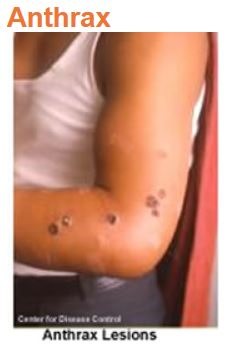